


ALOKA/HITACHI PROSOUND 3500SX COLOR 3D/4D DOPPLER ULTRASOUND SCANNER

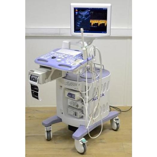
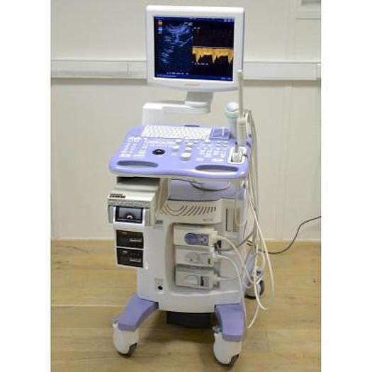
ALOKA/HITACHI PROSOUND 3500SX COLOR 3D/4D DOPPLER ULTRASOUND SCANNER
Fr14,280 (CHF)
or
Call + 41 (0)27 720 41 00
Description
with :
- 3D/4D abdominal volume probe
- 2D abdominal convex probe
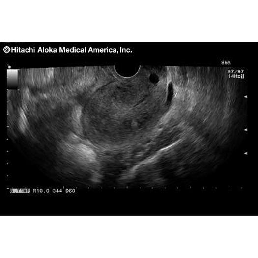
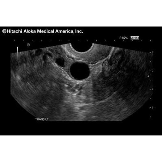
- 2D endovafgianle probe
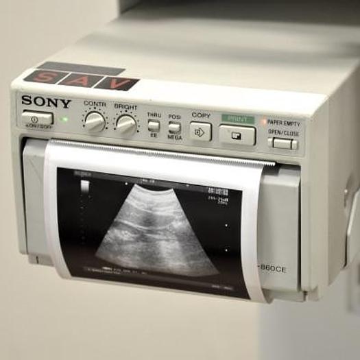
- Sony B&W printer
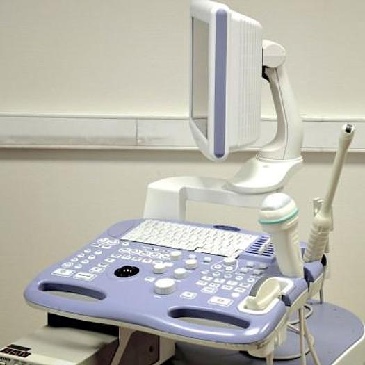
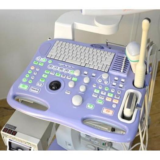
Ergonomics:
The compact, lightweight system is easy to move around.
Flicker-free monitor display reduces eye fatigue.
Control panel and display screen can be shifted up/down for your optimal position.
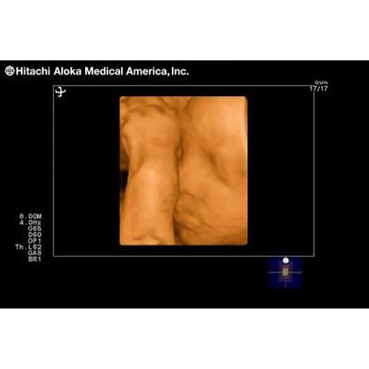
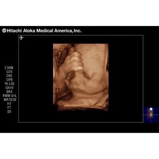
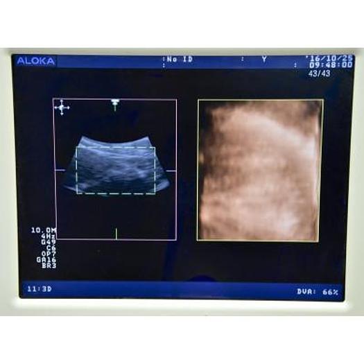
3D/4D imaging
Imaging of the fetus in motion, in real time by high-speed scanning.
The viewpoint of a 3D image can be freely rotated 360 degrees horizontally and vertically. Images you want to see, such as the face of a fetus, can easily be displayed independently of the position of the fetus. MPR (Multi Planar Reconstruction) allows the simultaneous display of three arbitrary orthogonal sections, enabling observation of the coronal section that is impossible to analyze with 2-D scanning. A single 4D probe is used for all routine Doppler, Color Flow and 4D examinations.
Real-time FAM (free agular M-mode)
Up to three M-mode cursors are individually moved and rotated during scanning for fast, accurate M-mode examination. For example, the cardiac function of a fetus can be easily checked regardless of its orientation.
Digital data and network management
The system supports DICOM 3.0. Patient and image data can be transmitted to the file server in the network.
The latest measurement data and ultrasound images can be quickly retrieved from the integrated hard disk.
Tissue Harmonic Echo (THE)
Tissue Harmonic Echo reduces defects caused by multiple near-field echoes and secondary lobes.
Quint Imaging Frequency (QFI)
QFI enables selection of optimum frequencies over an extremely wide range of the probe's bandwidth. It is possible to select higher resolution images or penetration imaging to suit the situation without changing the probe. QFI works not only on black & white images, but also on color flow images and Harmonic Echo images.
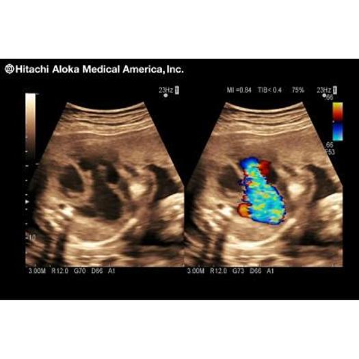
DDD (Dual Display Dynamic)
DDD is a function for displaying B-mode images with and Color Flow image simultaneously in real time. This function is useful for ultrasound-guided biopsy, as the examination can be carried out at high speed and facilitates differentiation of hypoechoic plaque, which is difficult to diagnose using B-mode images alone.
Specifications
| Condition | Used |
| Stock Number | 5164 |
| Pdf catalog | (click HERE) |