


ESAOTE MY LAB CLASS C ULTRASOUND SCANNER

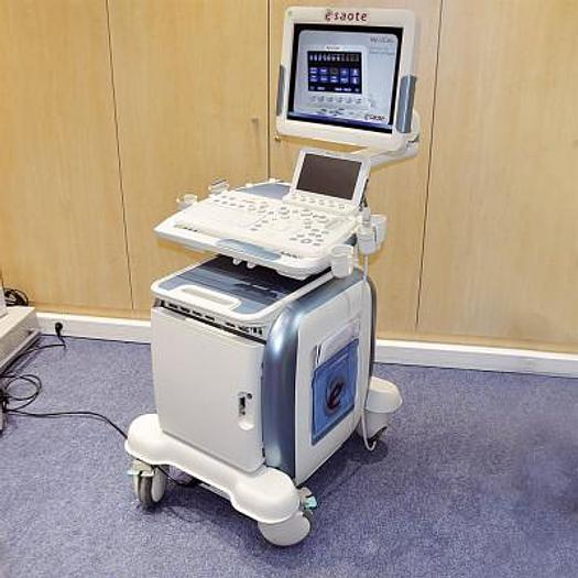
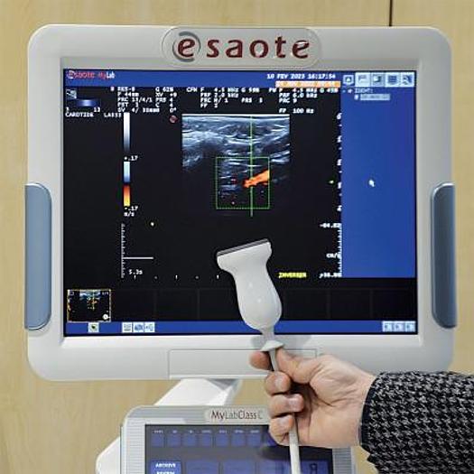
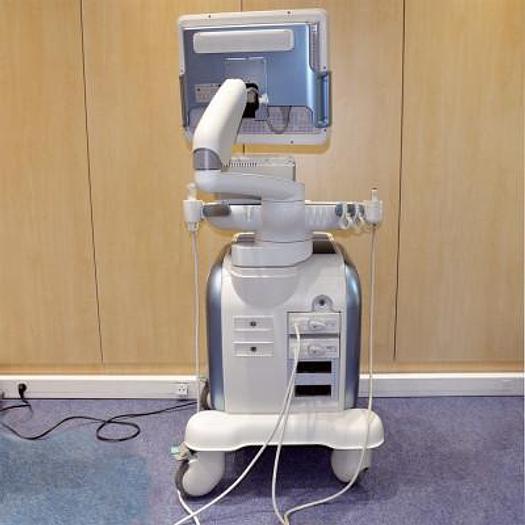
ESAOTE MY LAB CLASS C ULTRASOUND SCANNER
Fr11,550 (CHF)
or
Call + 41 (0)27 720 41 00
Description
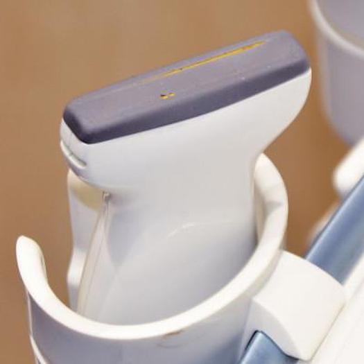
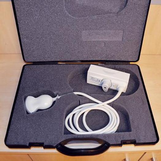
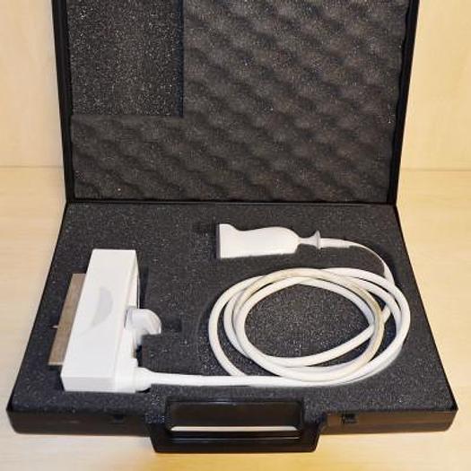
With 2 PROBES :
LINEAR PROBE 50MM 3.0 - 13. MHZ
CONVEX PROBE 50R 1.8-8.0 MHZ)
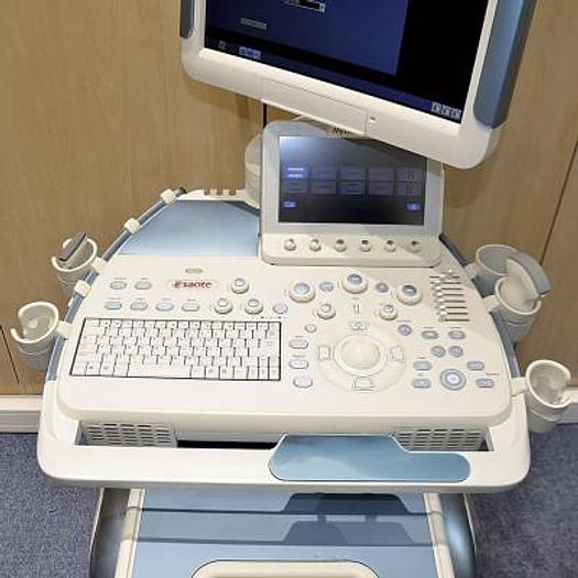
The innovative features of the MyLab™Class C allow the system to be placed in any type of environment, always providing easy, fast and reliable access to diagnostics and shared data.
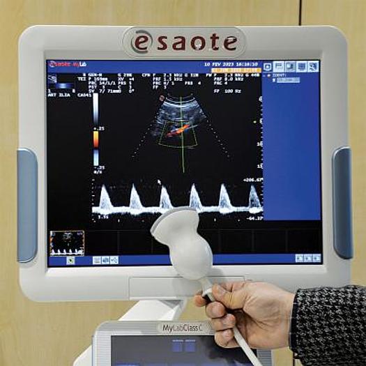
Prevention and quantification in cardiovascular imaging are accessible at your fingertips: the prevention suite with state-of-the-art RF-QIMT, RF-QAS, XStrain™, CFI technologies and iQProbes probes guarantee the best possible diagnostic approach. In general imaging today, CnTI™ (Contrast Tuned Imaging) for contrast agent procedures, HD CFM and XFlow, high-frequency imaging, X4D technology, Virtual Navigator and Elaxto elastosonography meet all clinical needs.
Click on the image to view images obtained with Esaote's cutting-edge technologies
Imaging processing: Esaote offers numerous technologies for image enhancement. With TEI™ , the harmonic signal is fully preserved without degrading acoustic information. MView and XView improve ultrasound image quality by reducing the presence of artifacts, background noise and superimposition.
CnTI™ - Adjusted contrast imaging for procedures with contrast agents: CnTI™, Esaote's revolutionary technology combined with the ultimate generation of ultrasound contrast agents delivers impressive clinical results due to the extremely accurate detection of microbubbles. The very low acoustic pressure applied increases the lifetime of the bubbles, with a view to clear identification of the arterial and late phase. The very high sensitivity of the probes and the low level of noise and artifacts enable precise diagnoses, both for the detection and characterization of lesions. A dedicated contrast quantification tool is also available.
HD CFM and XFlow - Extraordinary flow sensitivity and spatial resolution: The resolution and sensitivity of Color Doppler mode are of great importance for blood flow assessment, particularly when flow velocity and size are limited. HD CFM technology helps the user to define the right setting for maximum clinical information. For specific diagnostic processes where morphological information is more important than the hemodynamics themselves, XFlow produces images of great clarity with low artifacts, and less dependence on the insonation angle.
High-frequency imaging: Esaote's historic leadership in high-frequency imaging ensures an unexpected level of detail in applications where superficial images are required. 22 MHz transducers, XView, MView, ElaXto and X4D applications, and the "A Universe under the mm" software package are just a few examples of the technological potential of the MyLab™Class C system. The clinical results are simply astounding, as they open up new fields of research and take diagnosis to another level. Cutting-edge technologies such as ElaXto and X4D are implemented not as additional qualitative information, but as an important quantitative suite to produce an objective and rapid diagnosis.
X4D technology: The advanced 3D/4D software package benefits from innovations in the conventional visualization of 2D ultrasound images through spphisticated algorithms, and is capable of producing outstanding 3D/4D volume reconstructions. Measurements of length, area, perimeter, diameter and angle, as well as volume zones in a multi-dimensional display, enable quantitative analysis and qualitative acquisition, combined with a special database for archiving all personal data sets.
RFQIMT - RF-based qualitative measurement of media intima thickness for early detection of cardiovascular disease: RFQIMT technology aims to measure the thickness of a blood vessel in the area of the carotid artery selected for examination. Its ease of use, combined with real-time qualitative feedback, helps the operator to obtain accurate, reproducible results. Measurements (even if taken at different times during the examination) can be plotted in a standardized graph displayed together with reading indicators, helping physicians to establish their diagnoses and therapeutic procedures.
RFQAS - Qualitative measurement of arterial stiffness for early detection of cardiovascular disease: RFQAS technology measures the stiffness of a blood vessel in the area of the carotid artery selected for examination. Blood vessel wall stiffness is expressed in terms of brachial arterial pressure, with precise measurement of diameter and changes in diameter. Local blood pressure at the site of ultrasound measurements is also recorded. Local blood pressure and stiffness are derived as quantitative results based on sophisticated clinical studies.
Automatic measurement and adjustment: Quantifying the Doppler profile is an important issue in cardiology and vascular ultrasound examinations. Once the volume of interest has been determined and the Doppler trace is displayed on the monitor, the user can select real-time evaluation of all essential clinical parameters by activating the ADM function. Using the image freeze mode of your choice, you can also trace the Doppler contour and automatically record maximum, mean and minimum values. Features such as EF (Ejection Fraction) calculation and ADM (Automatic Measurement) provide quantification of the most important clinical parameters in the shortest possible time. This enables faster screening and precise patient management in cases of potential disorders, which can then be investigated further.
CFI (Coronary Flow Imaging) - Coronary Flow Imaging for the assessment of coronary artery blood flow and its main characteristics: The assessment of coronary blood flow characteristics is also significant with regard to baseline cardiac activity without any external induced cardiac stress. If a CFI Color Doppler preset is activated, the signal from coronary artery blood flow is optimized in relation to many of the concomitant blood flow velocity components present in the heart's atria and ventricles. The combination of the Cardiac iQ-probe and the CFI (Coronary Flow Imaging) preset offers superior performance in CFM/PW modes for coronary flow detection and measurement.
XStrain™: XStrain™ is a non-invasive tool that enables better exploration of myocardial function and quantification of aspects of heart physiology that were not possible to detect and quantify with previous ultrasound technologies. Myocardial velocity, myocardial overload and overload ratio enable early detection of cardiac function deficiencies (assessed as ejection fraction or stroke volume). Based on angle-independent technology, the XStrain™ function assesses the contractility of both left and right ventricles. XStrain™ is an innovative tool for the mechanical assessment of cardiac wall motion. It can therefore provide quantitative support for standard ultrasound examinations and be used to examine and monitor patients, in order to identify early signs of changes in cardiac wall motility.
Advanced Virtual Navigator tool for Fusion imaging (US and CT/MR/PET-CT): the Virtual Navigator tool with Fusion imaging applied to ultrasound enhances the information produced by an ultrasound scanner by combining it with a second imaging modality (CT, MR, PET or 3D US) in real time, offering all the advantages of different modalities in a single examination.
Virtual Biopsy - Advanced biopsy even in highly complex approaches: the Virtual Biopsy function enables you to follow a percutaneous procedure by superimposing needle tracking information on the real-time ultrasound image.
Specifications
| Condition | Used |
| Stock Number | 9213 |
| Date of manufacture | october 2017 |